Part of the “Rugby Proof Renegade” series – An extensive series of pre-habilitation programs aimed at injury reduction for the rugby athlete.
The knee is a commonly injured joint in rugby. The major problem with injuries to the knee is that they can often be very time costly leading to extended periods of time out of training and playing. A low grade ligament strain may only last a couple of days, a meniscal tear can lead to a couple of weeks out or something more serious like an ACL rupture will have you out of the game for 9 months or more. It’s for those reasons investing some time into injury prevention for the knee pays back big returns!
The knee is the articulation between your femur (thigh bone) and tibia (shin bone) supported in its role by the patella (knee cap). When most people think of the knee, they think of a hinge like joint that has two main movements: flexion – when the knee bends, and extension – when the knee straightens. Despite the simplistic view that it is a pure hinge joint moving in one plane of motion it actually has far more complex three-dimensional capabilities. As such it is important to train the knee to be able to control all of these movements.
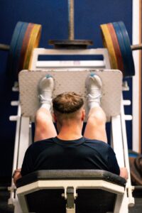
Major muscle groups acting on the knee are the quads, hamstrings and calf but the adductors play a role and the muscles of the hip also indirectly control it, as do the muscles of the trunk, foot and ankle. It is important to ensure that these muscle groups are just as able to contribute to the control of the knee. Make sure you take a look at the articles on rugby proof ankle and core stability to maximize the injury prevention program for the knee. You’ll see that a lot of the example exercises shown in this article involve the ankle, the hip as well as the whole body. This is because the transfer of the exercises to the rugby field is increased when they include multiple joints and muscles working together. It is possible to damage all of the muscles around the knee but the management of these injuries will be addressed in the information on muscle strains and tears.
Unsurprisingly the knee is most commonly injured during contact situations when the knee has to manage large forces across the joint. A wide range of injuries with a wide range of recovery times can occur and we will look at some of the more common injuries when we address the anatomical structures of the knee. Non contact injuries and overuse injuries also occur and generally require a different approach of management and rehabilitation. On the rugby field, tackles side steps, rucks and mauls can put you in potentially risky situations. This is part of the game and there will always be risks on a rugby field. It’s part of why we love the game. We would always take the approach that we want our rugby players to be strong, we know they’ll never be 100% safe. If they were 100% safe, they wouldn’t be on a rugby field where they belong. If you want more information then take a look at our information on injury prevention for rugby.
First we will take a look at the anatomy of the knee so you can better understand what happens when you injure your knee.
Anatomy of the Knee
The knee is made up of a number of structures that you may have heard of before. Here are some of the key anatomical structures of the knee and their functions:
The bones:
The femur (thigh bone). The base of the longest bone in the body, the femur, forms the top of the knee joint. Its curved shape at the end of the femur forms two femoral condyles that articulate with the tibia (shin bone) below. These femoral condyles are covered in articular cartilage that will be discussed more when we look at joint surface injuries.
The tibia (shin bone): To compliment the convex curve of the femur the tibia has more of a convex shape that is further enhanced by the meniscii (cartilage) that will be presented in the soft tissue section of the article.
The patella (knee cap): The kneecap sits in-between the groves of the two condyles at the base of the femur. Its role is to help distribute forces from the thigh to the shin bone through the pull of the quadriceps muscles that are found on the front of the thigh.
The Knee Ligaments
Strong cord like structures that provide some mechanical stability to the joint but that also provide a huge amount of proprioceptive and joint sense information to the brain. They nearly always work in combination with the muscles that surround the joint where they are located.
Anterior Cruciate Ligament (ACL):
The one no rugby player wants to injure. The ACL is found deep in the knee joint and anatomically it serves to limit forward translation of the tibia in relation to the femur. It works closely with the hamstrings that prevent the same movement when they contract. It can be vulnerable during side stepping and contact situations. A complete rupture of the ACL leads to a prolonged time off the field with recovery for both surgical and non-surgical recovery taking months to complete, often nine months or more. It used to be the case that most complete ruptures of the ACL were surgically repaired in sporting populations but good results have also been seen through conservative (non-surgical) management.
Posterior Cruciate Ligament (PCL):
The PCL sits slightly behind the ACL and crosses the back of the joint in a close to vertical direction. Its purpose is the opposite of that of the ACL. The PCL prevents backwards translation of the tibia relative to the femur. Because of this function it works closely with the quadriceps muscles. The PCL can be vulnerable to contact based injuries when the shin bone is forced backwards, or when the knee hyperextends.
Medial Collateral Ligament (MCL):
The MCL sits on the inside of the knee. It is a broad ligament that protects the knee from lateral forces applied to the outside of the knee. It is vulnerable to injury when contact is made with the outside of the knee, especially when the foot is planted. Injury to this ligament is one of the most common knee injuries for rugby players and can cause a wide range of periods out of the game depending on the grade of damage to the ligament from a matter of days up to twelve weeks for a high grade injury. Most injuries heal without surgery but may require use of a brace to protect the healing ligament. If surgery is needed the recovery can take more than nine months.
Lateral Collateral Ligament (LCL):
The LCL is on the opposite side of the knee, the lateral side. It is thinner than the MCL because as a general rule it deals with less force than the MCL. The LCL protects the knee from lateral forces applied to the inside of the knee joint that could open or gap the joint on the outside of the body. Because there is often the opposite leg and hip in the way of the inside of the knee LCL injuries are less common than MCL injuries but they can occur and often occur in combination injuries with the other ligaments of the knee when a large amount of force goes through the knee joint.
Cartilage
Meniscii: The meniscii sit in the space between the shin bone and the femur. You have on on the inside of your knee, the medial meniscus. And one on the outside of your knee, the lateral meniscus. These two structures serve to cushion the compressive forces that the knee is exposed to during weight bearing activities. The cartilage discs can become worn and this is a natural process of aging in most cases. More acute injuries can occur when the knee is rotated under load when the foot is plated in the ground. Meniscal injuries along with MCL injuries discussed above are one of the most common injuries to the knee for a rugby player. Tears of the meniscii are common and can occur in both the medial and lateral meniscus. It is more common in the medial meniscus. These injuries can recover on their own but sometimes they will require surgery to either trim the meniscus or to repair the meniscus with stitching. If the injury requires trimming the recovery time on average is around 4-6 weeks. If the meniscus is removed it can take up to 3-4 months.
The Unhappy Triad of the Knee
In unfortunate cases it is possible to injure a number of structures in the same knee at the same time. One such example is known as the unhappy triad where the ACL, MCL and medial meniscus all get injured at the same time. Due to the biomechanics of the knee the classic ACL mechanism of injury where the knee is under load, flexed and rotated all three structures are susceptible to injury.
Cartilage and Osteochondral Injuries
Osteochondral injuries are injuries that involve the smooth cartilage that covers the ends of bones and the bone itself that sits underneath the cartilage. These injuries can be extremely problematic for rugby players. There is a large range of osteochondral injuries ranging from a small crack in the cartilage only, to a larger hole in the cartilage that exposes the underlying bone or a very advanced injury would be removal of the cartilage and significant damage to the underlying bone as well. The main issue with these injuries is that they have very limited capacity to heal themselves so what starts out as a small tear or gap in the cartilage can progress to a more significant injury as time goes by and as training and matches continue.
Management can be tricky. If fragments of the bone or cartilage remain in the knee joint they can cause the knee to become unstable, cause it to lock in a position if the fragments get trapped in the joint or cause the joint to become very inflammatory and reactive. In these cases surgery may be required to remove the fragments.
For a stable non-reactive knee – load monitoring and paying attention to symptoms is crucial. If the knee becomes sore or inflamed it’s an indicator the levels of loading are too high and need to be adjusted. If the load tolerance of the knee becomes that limited advanced surgical cartilage transplant and microfracture techniques may be the only option. Recovery time from these procedures is significant, 12 months plus so it is often seen as a last resort, especially in high level sports.
Osteoarthritis (OA)
OA is a very common knee complaint and is a condition that describes the general wear and tear of the joint that occurs as we age. Occasionally activity, injury and medical interventions can accelerate the rate of wear in our cartilage. Degenerative changes have been shown to be very common in knees as we age and signs on scans do not correlate well with clinical symptoms. So always pay attention to how the knee actually feels. OA can appear between the tibia and the femur as well as the femur and the patella.
Mechanical and Overuse injuries
The next two injuries, patella tendon and patellofemoral joint conditions can be caused by a number of factors like the strength of the quads, the position and the anatomy of the bones of the knee, the control and position of the hip as well as the position of the tibia and the strength and control of the foot and ankle. As such management can be tricky and treating just the site of pain can often lead to poor outcomes. What we will present here is a brief introduction to the conditions. Our injury prevention approach is a great way to prevent these conditions but if you feel like you suffer from either get checked out by a professional to get on to the best rehab pathway for your condition. They both present with pain at the front of the knee but the management of each is very different. Don’t get caught chasing the wrong rabbit.
Patella Tendon Issues
The most common knee tendon issue in the knee is the patella tendon. Pain and soreness in the patella tendon at the front of the knee is a relatively common complaint, especially in jumping athletes. That’s why it is commonly referred to as “jumper’s knee”.
Signs and symptoms include pain on the tendon itself, pain on the either or both of the insertions of the tendon to the patella and the tibia. Initially just soreness after training sessions that resolves after a brief rest. This can progress to soreness at the start of training sessions that eases as the tendon gets warmed up but becomes sore again following the session. The next stage of the condition would be to consistently feel pain in the tendon, even when performing low level activities like walking or unloaded squats.
Like a lot of tendon injuries they typically manifest when the loading of the tendon exceeds its capacity to deal with that load. Over time the tendon gets trapped in a downward cycle of getting loaded during training and matches, not having enough time to recover, and getting loaded again. This cycle of loading and inadequate recovery creates inflammation and soreness. With these injuries appropriate load modification and rehab is needed. Without it the cycle continues and symptoms often continue to get worse. Rehab often focuses on strengthening the quads and the tendon itself using techniques, exercises and systems to optimise the loading of the tendon and most importantly allowing it to adapt to increasing loads over time. In advanced cases injection therapies like Platelet Rich Plasma (PRP) injections may be considered but this should be a late stage option and should not be considered before a well constructed rehab program has been put in place and followed. In very rare cases surgery may even be considered.
Patellofemoral Joint (PFJ) Pain or Stress Syndrome
PFJ pain occurs when the posterior (back) surface of patella rubs against the surface of the femoral condyles.
Signs and symptoms include soreness at the front of the knee during and after activity that generally gets worse as the activity continues. Pain on sitting for long periods is also a common sign.
Management focuses on improving the alignment of the knee joint focusing on the hip, knee and foot and ankle in any combination depending on which is the main contributing factor. Orthotics may be considered if the position and control of the foot and ankle is a driver of the pain. Complete pain resolution can be difficult but maintaining a healthy weight and an appropriate training program can go a long way to reducing pain and improving function.
If you have any questions about the information contained in this article then please contact us through the website or our social media channels.
Signs of serious injury
If you have any concerns at all about an injury the first thing you should do is get it assessed properly. You can use our online physio service to do this
Key signs of a serious knee injury include:
Reduced ability to put your body weight through your knee
A sensation of instability or giving way
Reduced range of movement in any range of motion
Significant swelling
A deformity or your knee not looking right
New and additional sounds when you move your knee – clicking or clunking from inside the joint
If you have any of these signs or symptoms then get your knee assessed as soon as possible.
Take a look at our complete list of injury prevention exercises for the knee on our YouTube channel. We are always adding to our library so make sure you subscribe to stay up to date with all of our content!